|
Last week we saw an overview of the gait cycle. The basic phases in gait cycle and the fundamental components of the stance and swing phases. The subsequent week’s post will consist of elaborating the biomechanics of the various joints in the lower extremities and the trunk.
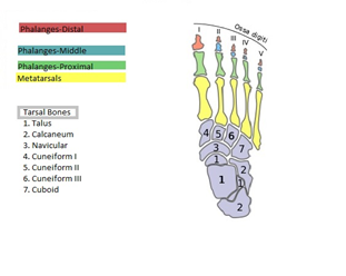
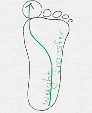
We will consider the body from the ground up or from the foot to head. The foot is made up of 26 bones and 33 joints! Though the amazing fact is it works flawlessly as one unit. While it will be boring to consider all the joint movements, it is good to know that the foot moves in multiple planes ensuring smooth transfer of weight from the heel (calcaneum) to the toes (phalanges). The major foot movements include lifting up at heel strike, pushing back at push-off phase, as well as the subtle rotational motion (pronation-supination) during weight transference from heel to toes. The ankle joint is formed by the two lower leg bones (tibia and fibula) with the foremost tarsal (seven tarsal bones in all) bone called the talus. The two leg bones form an inverted cup shaped cavity which is deep and houses the head of talus. This cavity is also known as the ankle mortise and is responsible for the stability of the ankle joint. The ankle joint is a hinge joint and the only movement possible here is upward (dorsiflexion as in heel strike) and downward (plantarflexion as in push-off) movement. The side to side and rotational movement during weight transfer occurs at the tarsal and metatarsal joints. The heel bone or the calcaneum is the largest bone in the foot. At heel strike the heel bone is everted, that is the heel lands on the outer edge. This is the initial impact of landing at heel strike. The initial impact is absorbed by pronation in the foot when the talus rotates antero-medially allowing the leg bones (tibia and fibula) to come into weight bearing position, to transfer the entire body weight to the foot. The anterior leg muscles like the tibialis anterior, the extensor hallucis longus, and extensor digitorum slowly lengthen permitting smooth lowering of the foot to the ground. As the knee flexes and weight is being transferred to the foot, it initially pronates or winds for impact absorption and then unwinds or supinates to allow maximal contact of foot with the ground (early stance). The transverse tarsal joints (talo-navicular and calcaneo-cuboid) are loose and accommodate the foot to uneven terrain. At mid stance, the body moves over a stable foot and the weight gradually shifts to the forefoot from tarsals to the five ray-like metatarsal bones (mostly 1st-3rd metatarsal rays). Here the arches with their muscular and ligamentous support are very important for weight transfer. The posterior leg muscles like the tibialis posterior and the peroneus longus are key muscles locking the transverse tarsal joints hence making the foot into a rigid lever. The foot continues supination as the heel lifts off the ground and the ball of the foot (1st metatarsal head) bears most load during the end of push-off phase. The Achilles tendon, which initially lengthens, now has sufficient potential energy to propel the body forward with a strong concentric contraction. Amazing isn’t it? The subtle movements in the joints of the foot, the display of combination of stability as well as mobility and the impeccable timing with which this is executed (considering average walking speed of adult is 100-115 steps/minute!) [For ease of understanding, here is a list of the bones in the foot: 7 tarsal bones: talus, calcaneum, navicular, cuboid and the 3 cuneiform bones 5 metatarsal bones: five rays one of each leading to the corresponding toe 14 phalanges: 2 in the big toe and 3 each from the 2nd to 5th toe.]
0 Comments
Your comment will be posted after it is approved.
Leave a Reply. |
Details
AuthorAmi Gandhi is a licensed physical therapist in the state of California. She is the owner of StableMovement Physical Therapy, a small boutique practice in San Jose that offers patient centered, one-on-one, hands-on physical therapy. Archives
March 2018
Categories |

